OSTEOPOROSIS – GOALS – FACTS – ADVANCED MEDICATIONS

BONE LOSS IS FASTER THAN THE GAIN 

*PREVENTABLE – (YES) *TREATABLE – (YES)

Goals: From NOF National Osteoporosis Foundation*

Reduced Trabecular network
  • Greater awareness about the importance of bone health, its impact on patients and caregivers, and cost-effective steps that can be taken to optimize bone health, prevent fractures and improve health outcomes.
  • End the stigma and ageism associated with osteoporosis to assure that bone health gets the high priority and attention it deserves. 
  • Everyone deserves access to affordable, high-quality, age-appropriate bone health screening.
  • Access to effective therapies to treat osteoporosis should be affordable and determined by each patient and their health professional.
  • Best practices in care management and coordination for those who have suffered bone fractures should be replicated and appropriately incentivized.

KNOWN FACTS

ABOUT OSTEOPOROSIS
Osteoporosis is a disease of the bone.
Osteoporosis is a “silent disease” until it is complicated by fractures, because you cannot feel your bones getting weaker, lighter and porous.
Osteoporosis is common.

INCIDENCE

Statistics from the International Osteoporosis Foundation, worldwide, 1 in 3 women over the age of 50 years and 1 in 5 men will experience osteoporotic fractures in their lifetime.
• Approximately 10 million Americans have osteoporosis and another 44 million have low bone density, living with increased risk.
• This means that half of all adults age 50 and older are at risk of breaking a bone and should be concerned about bone health.
• The disease is responsible for an estimated two million broken bones per year, yet nearly 80 percent of older Americans who suffer bone breaks are not tested or treated for osteoporosis.
• One in two women and up to one in four men over age 50 will break a bone due to osteoporosis.
• People with osteoporosis can break a bone from a minor fall, or in serious cases, even from simple actions like sneezing or bumping into furniture.

Osteoporosis is serious, even deadly.
• A woman’s risk of fracture is equal to her combined risk of breast, uterine and ovarian cancer.
• A man is more likely to break a bone due to osteoporosis than he is to get prostate cancer.
• 24 percent of hip fracture patients age 50 and over die in the year following the fracture.
• Every year, of nearly 300,000 hip fracture patients, one-quarter end up in nursing homes and half never regain previous function.
Osteoporosis is costly.
• Osteoporosis-related bone breaks cost patients, their families and the healthcare system
• By 2025, experts predict that osteoporosis will be responsible for three million fractures resulting in $25.3 billion in costs.
Osteoporosis is preventable.
• About 85-90 percent of adult bone mass is acquired by age 18 in girls and 20 in boys. NOF is currently updating a scientific manuscript regarding Peak Bone Mass and how people can build and maintain strong bones throughout their lifespan.
• Building strong bones during childhood and adolescence can help prevent osteoporosis later in life.
Osteoporosis is manageable.
Healthy diet and Regular Exercise can help slow or stop the loss of bone mass and help prevent fractures.
• About half of osteoporosis-related repeat fractures can be prevented with appropriate treatment.

This image has an empty alt attribute; its file name is capture.jpg
Osteopenia apparent on plain X-Ray

INVESTIGSTIONS FOR OSTEOPOROSIS
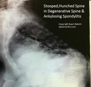
PLAIN X-RAYS

Plain x-rays esp. the wrist, spine and hips give a fair assessment of osteopenia and frank osteoporosis, if noticed, as most physicians mostly lay less importance to this fact.

BLOOD TESTS CONSISTING OF

Basic: Serum Calcium, Phosphorus, Alkaline Phosphatase

TSH and PTH if any abnormal findings for Calcium , Phosphorus and alkaline phosphatase

Advanced: Bone Markers: bone markers can be useful in detecting the imbalance and bone loss.

At least 1 formation marker and 1 resorption marker from the list

List of bone formation markers

  • Serum total alkaline phosphatase
  • Serum bone–specific alkaline phosphatase
  • Serum osteocalcin
  • Serum type 1 procollagen (C-terminal/N-terminal): C1NP or P1NP

List of bone resorption markers

  • Urinary hydroxyproline
  • Urinary total pyridinoline (PYD)
  • Urinary free deoxypyridinoline (DPD)
  • Urinary collagen type 1 cross-linked N-telopeptide (NTX)
  • Urinary or serum collagen type 1 cross-linked C-telopeptide (CTX)
  • Bone sialoprotein (BSP)
  • Tartrate-resistant acid phosphatase 5b

DEXA* – A bone density test is the best way to diagnose osteoporosis and determine a treatment plan. If your T-score is -2.5 or lower, indicating that you have osteoporosis, or if you have other significant risk factors for breaking a bone, discuss with your doctor about starting an osteoporosis planned tailored treatment plan including medication.
• Discuss the risks and benefits of all medication in the treatment.
• Take recommended amount of calcium and vitamin D daily from foods and/or supplements.
• DEXA should be repeated at least every two years to monitor treatment. After starting a new osteoporosis medicine, many healthcare providers will repeat a bone density test after one year.

*DEXA – Dual-energy X-ray absorptiometry

OSTEOPOROSIS ADVANCED MEDICATIONS OPTIONS AVAILABLE 

  1. FOSAMAX 70 MG 

ONCE PER WEEK, for a year and then repeat DEXA SCAN  

Mostly very effective, simple and safe, fewer side effects, insurance covered cost effective

2. IBANDRONATE  

3 MONTHS – BONVIVA

Prefilled syringes are for single use only. The recommended dose of BONIVA Injection for the treatment of postmenopausal osteoporosis is 3 mg every 3 months administered intravenously over a period of 15 to 30 seconds. 

INDICATIVE PRICE 192/-AED approx 

4. PROLIA (DENOSUMAB )

ONCE IN 6 MONTHS 

60 MG, S/C injection,

INDICATIVE PRICE 1180/- aed approx 

5. ZOMETA 1 YEAR 

zoledronic acid for inj 

Reclast (zoledronic acid) is a bisphosphonate used to treat Paget’s disease, high blood levels of calcium caused by cancer (hypercalcemia of malignancy, multiple myeloma (a type of bone marrow cancer) or cancer that has spread from elsewhere in the body to the bone, to treat or prevent osteoporosis in postmenopausal 

INDICATIVE PRICE 866/- AED 

6. FORTEO 

DAILY TERIPERIIDE Inject FORTEO in the thigh or abdomen once daily with the easy-to-use FORTEO delivery device 

INDICATIVE PRICE – NA 

Patients taking Actonel showed a bone mineral density (BMD) increase of 2.7%, while those taking the Reclast showed increased BMD of over 4%. 

REFERENCES

*courtesy from NOF with due permission and sincere thanks, for all the quotes and links, for wider dissemination of information on Osteoporosis

2. Eur J Rheumatol. 2017 Mar; 4(1): 46–56.
Published online 2016 Dec 30. doi: 10.5152/eurjrheum.2016.048
PMCID: PMC5335887
PMID: 28293453
An overview and management of osteoporosis
Tümay Sözen,1 Lale Özışık,2 and Nursel Çalık Başaran2

BONE & CARTILAGE HEALTH RECIPES

NUTRITIOUS IDLY ( Rice & Lentil Cake cooked in Steam for Breakfast ) ENRICHED WITH VITAMIN D WITH SOYA AND SPICES

Ingredients 

Idli Dosa Batter Fresh or Packaged

Soya nuggets

Green chilly chopped fine

Ginger in thin slivers 

Red paprika 

Pudina Parantha masala – Mixed Spice with dried Mint leaves

Fresh grounded Black peppers 

Chopped coriander leaves 

Chopped Onion and Tomato 🍅Optional 

Just Mix and keep in room temperature up-to an hour if time permits or ready to use

Routine Idli cooking 

Recipe for Bone and Cartilage Health Series 

NUTRITIOUS OMELETTE WITH SOYA NUGGETS & SPICES ENRICHED WITH VITAMIN D

Omega 3 eggs

Green chilly chopped fine

Ginger in thin slivers 

Red paprika 

Pudina Parantha masala 

Fresh grounded Black peppers 

Chopped coriander leaves 

Chopped Onion and Tomato 🍅Optional 

Cook on very low flame COVERED and one side

Copyright Dr Kapil Bakshi <bakshiortho.com>

Recipe for Bone and Cartilage Health Series 

MUSCULO-SKELETAL ULTRASOUND ‘MSK’

Dr Bakshi has completed the ‘MSK Ultrasound Course and Hands on training’ from the worlds premier Radiology & Ultrasound training institute at Thomas Jefferson University Hospital, Philadelphia this October, 2016 to refine and update current knowledge and technology used.

Dr. Bakshi is one of the few Orthopedic Surgeons who do their own Ultrasound for the diagnosis, while treating the pathology with precision ultrasound guided Local and Intra-articular injections and Regenerative Therapies in sterile operative conditions.

MSK ultrasound saves time as it is done as an office procedure, many times the pathology is so apparent that an MRI is often saved, saving further costs of treatment to the insurance companies and cash paying patients.

Ultrsound is safe and has no ionizing radiation.

Further the scan is done for the area of interest according to the clinical findings seen and felt by the treating surgeon, these are then visualized by the ultrasound. Unwanted scanning of normal areas is not done saving time both for the patient and the doctor. The few radiologists who have the skills of doing soft tissue  MSK ultrasound scan the entire area as they are not aware of where the pathology is.

Advantage over MRI

a. Dynamic ‘Real time’ ultrasound – motion of joint, muscle and tendon is evaluated ‘live’ with the ultrasound with the ensuing motion.

b.Stress test evaluation of ligament injuries.

c. Floating debris and non opaque foreign bodies in an effusion.

In a short span of time we will introduce ultrasound guided percutaneous surgery with a 2-3 mm incision using micro endoscopic surgical knife and ultrasound guided Hypodermic needle surgeries.

msk-ultrasound-course-certificatemusculoskeletal-ultrasound-course-october-7-8-2016-certificate-kapil-bakshimusculoskeletal-ultrasound-course-october-7-8-2016-certificate-kapil-bakshi
SHOULDER EXAMINATION with Ultrasound – Procedure in brief

The long head of the biceps tendon – forearm in supination and resting on the thigh or with the arm in slight external rotation. The tendon is examined in a transverse plane (short axis), where it emerges from under the acromion, to the musculo- tendinous junction.

Subscapularis tendon, the elbow remains at the side while the arm is placed in external rotation. The subscapularis is imaged from the musculotendinous junction to the insertion on the lesser tuberosity. Dynamic evaluation – patient moves from internal to external rotation.

Supraspinatus tendon, the arm can be extended posteriorly, and the palmar aspect of the hand can be placed against the superior aspect of the iliac wing with the elbow flexed and directed toward the midline (instruct the patient to place the hand in the back pocket. Transducer

Posterior aspect of the infraspinatus and teres minor tendons should be examined by placing the transducer at the level of the glenohumeral joint below the scapular spine while the forearm rests on the thigh with the hand supinated. Internal and external rotation. Visualize the teres minor tendon, the medial edge of the probe should be angled slightly inferiorly.

Rotator cuff, the cuff should be compressed with the transducer to detect nonretracted tears. Contralateral side evaluation useful. Dynamic evaluation – assess the cuff tear extent. In patients with a rotator cuff tear, the supraspinatus, infraspinatus, and teres minor – examined for atrophy.

Subacromial-subdeltoid – bursal thickening or fluid.

Glenohumeral joint with the probe placed in the transverse plane from a posterior approach to evaluate for effusions, intra-articular loose bodies, synovitis, or bony abnormalities. Suprascapular notch and spinoglenoid notch also may be evaluated.
Acromioclavicular joint should be evaluated with the probe placed at the apex of the shoulder, bridging the acromion and distal clavicle.1–3


Rare Surgeries and Rare Case Studies

Median Nerve Neurofibroma

image
Median Nerve Neurofibroma

Severe clinical signs of Carpal Tunnel Compression Syndrome CTCS were seen in this young lady. There were no other visible or symptomatic masses seen over the body.

On exposing the mass, this inoperable median nerve lesion was seen, dissected and compression was relieved. Few fibers from the ulnar side were taken for a biopsy  which confirmed the nerve lesion. The bulging nerve could be covered with the stretched out skin. The wound healed well primarily and neurological symptoms were relieved. There were no distinguishable clinical effects of the biopsy.

The patient was symptom-free for two years till she was lost to follow-up.

Ankylosing Spondylitis

Ankylosing Spondylitis is a variant of arthritis that affects the spinal column, pelvis and eyes. It’s symptoms include pain and stiffness from the neck down to the lower back up to the coccyx (tail bone). The spinal bones (vertebrae) may fuse together, resulting in a rigid inflexible spine. These changes could be mild or severe, and may lead to a bent stooped-over hunching posture gradually over time. Early diagnosis and supervised management helps control pain and stiffness and may decrease or prevent further revealing deformity.

Incidence of Ankylosing Spondylitis

Ankylosing spondylitis affects men more than women in about 0.1% to 0.6% of the adult population. Although it can occur at any age, spondylitis most often affects men in their adolescence and 20s. It is less common and has lesser symptoms in women.

Symptoms of Ankylosing Spondylitis

The most common and early symptoms include:

  • Chronic pain and stiffness in the lower back, buttocks and hip areas that continue for more than two or more months esp. in the mornings. Spondylitis often starts around the sacro-iliac joints, where the sacrum joins the ilium bone of the pelvis. This extends to the upper spine, tendon and ligament attachments.
  • Bony fusion. It can cause an ‘fusion’ of the bones, which may lead to abnormal joining of bones when attempted healing occurs with calcification of vertebral ligaments. Fusion affecting bones of the neck, back, or hips  areas may restrict a person’s ability to perform Activities of Daily Living (ADL). Fusion of the ribs to the spine and/or breastbone may limit chest expansion and affect breathing.
  • Pain in ligaments and tendons. Spondylitis also may affect some of the ligaments and tendons. Achilles Tendonitis (inflamed tendon) may cause stiffness in the heel area. Other ligaments get involved gradually. Ankylosing &amp; Degenerative Arthritis
  • Compression Fractures. Weak crumbling vertebral bones increases hunching of back.
  • Late complications include Eye pain, photophobia and blurring (Uveitis). Heart dysfunction when aorta is involved in the inflammation.
  • Fever fatigue and appetite loss could be associated.

Causes

The specific cause of ankylosing spondylitis is unknown, a strong genetic  factor or family link may be involved. Some people with spondylitis carry a gene called HLA-B27 and people carrying this gene are more likelier to develop spondylitis. Up to 10% of people with HLA-B27  have no signs and symptoms.

Test and Diagnosis

  • Physical Examination: Range of spine flexibility (Serial checks)
  • Measurements of the chest expansion (Serial checks)
  • X-Rays of the back and pelvis (Oblique views of sacro-iliac joints)
  • Lab tests to check the level of inflammation CRP and ESR with routine blood counts
  • To rule out other arthritis in early stages including RA test and Anti CCP test
  • Blood test for HLA-B27

Treatment and Medication

Ankylosing spondylitis is incurable, but there are treatments that can reduce alleviate pain and improve function being the goals of treatment. Recommended to maintain a good posture, prevent deformity, and preserve the ability to perform normal activities of daily living (ADL). A team approach to treat spondylitis is recommended including the patient, doctor, physical therapist, and occupational therapist. In patients with severe fixed deformities, osteotomy and fusion may be required.

A program of daily exercises helps reduce stiffness, strengthen the muscles around the joints and prevent or minimize the risk of disability and deformity.

Deep breathing exercises may help keep the rib cage flexible.

Swimming is highly recommended.

Certain drugs help provide relief from pain and stiffness, and allow patients to perform their exercises with minimal discomfort. In moderate to severe cases, other drugs may be added to the treatment regimen.

Level 2: DMA (Disease Modifying Agents) such as methotrexate can be used when routine drugs alone are not enough to reduce the pain, stiffness and inflammation. These drugs have more toxicity and serial blood checks are required along this line of treatment.

New Drugs called biologics : Certolizumab pegol (Cimzia), (Humira) Adalimumab, Etanercept (Enbrel), golimumab (Simponi), and Infliximab (Remicade) — have been FDA-approved. Steroid injections into the joint  may be helpful in some cases. Antidepressant can be prescribed.

Above listed TNF blockers can reactivate latent TB (tuberculosis) and neurological conditions.

Artificial joint replacement surgery may be a treatment option in some patients with advanced joint stiffness affecting knees and hips.

Management of each patient is tailor-made


PATIENT TESTIMONIALS