Dr Bakshi has completed the ‘MSK Ultrasound Course and Hands on training’ from the worlds premier Radiology & Ultrasound training institute at Thomas Jefferson University Hospital, Philadelphia this October, 2016 to refine and update current knowledge and technology used.
Dr. Bakshi is one of the few Orthopedic Surgeons who do their own Ultrasound for the diagnosis, while treating the pathology with precision ultrasound guided Local and Intra-articular injections and Regenerative Therapies in sterile operative conditions.
MSK ultrasound saves time as it is done as an office procedure, many times the pathology is so apparent that an MRI is often saved, saving further costs of treatment to the insurance companies and cash paying patients.
Ultrsound is safe and has no ionizing radiation.
Further the scan is done for the area of interest according to the clinical findings seen and felt by the treating surgeon, these are then visualized by the ultrasound. Unwanted scanning of normal areas is not done saving time both for the patient and the doctor. The few radiologists who have the skills of doing soft tissue MSK ultrasound scan the entire area as they are not aware of where the pathology is.
Advantage over MRI
a. Dynamic ‘Real time’ ultrasound – motion of joint, muscle and tendon is evaluated ‘live’ with the ultrasound with the ensuing motion.
b.Stress test evaluation of ligament injuries.
c. Floating debris and non opaque foreign bodies in an effusion.
In a short span of time we will introduce ultrasound guided percutaneous surgery with a 2-3 mm incision using micro endoscopic surgical knife and ultrasound guided Hypodermic needle surgeries.
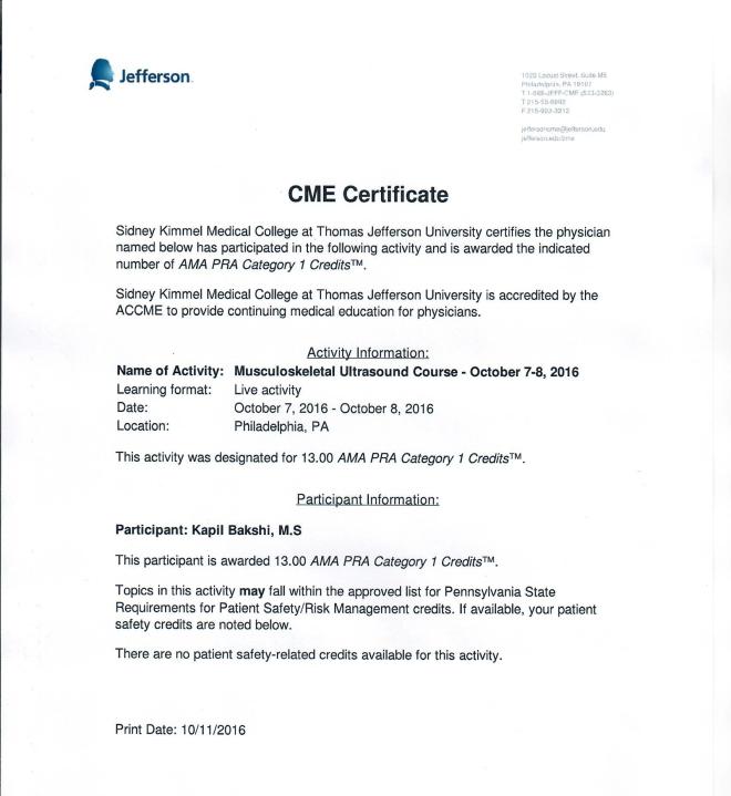 musculoskeletal-ultrasound-course-october-7-8-2016-certificate-kapil-bakshimusculoskeletal-ultrasound-course-october-7-8-2016-certificate-kapil-bakshi
SHOULDER EXAMINATION with Ultrasound – Procedure in brief
The long head of the biceps tendon – forearm in supination and resting on the thigh or with the arm in slight external rotation. The tendon is examined in a transverse plane (short axis), where it emerges from under the acromion, to the musculo- tendinous junction.
Subscapularis tendon, the elbow remains at the side while the arm is placed in external rotation. The subscapularis is imaged from the musculotendinous junction to the insertion on the lesser tuberosity. Dynamic evaluation – patient moves from internal to external rotation.
Supraspinatus tendon, the arm can be extended posteriorly, and the palmar aspect of the hand can be placed against the superior aspect of the iliac wing with the elbow flexed and directed toward the midline (instruct the patient to place the hand in the back pocket. Transducer
Posterior aspect of the infraspinatus and teres minor tendons should be examined by placing the transducer at the level of the glenohumeral joint below the scapular spine while the forearm rests on the thigh with the hand supinated. Internal and external rotation. Visualize the teres minor tendon, the medial edge of the probe should be angled slightly inferiorly.
Rotator cuff, the cuff should be compressed with the transducer to detect nonretracted tears. Contralateral side evaluation useful. Dynamic evaluation – assess the cuff tear extent. In patients with a rotator cuff tear, the supraspinatus, infraspinatus, and teres minor – examined for atrophy.
Subacromial-subdeltoid – bursal thickening or fluid.
Glenohumeral joint with the probe placed in the transverse plane from a posterior approach to evaluate for effusions, intra-articular loose bodies, synovitis, or bony abnormalities. Suprascapular notch and spinoglenoid notch also may be evaluated.
Acromioclavicular joint should be evaluated with the probe placed at the apex of the shoulder, bridging the acromion and distal clavicle.1–3
